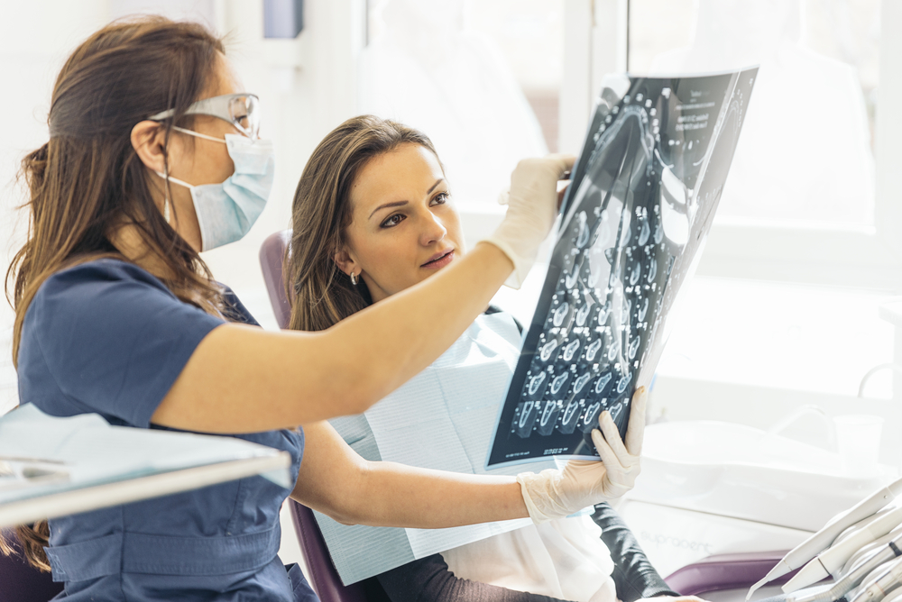
Dental radiographs, commonly known as X-rays, play a crucial role in modern dentistry and dental laboratory work. They provide detailed images of teeth, gums, and surrounding structures, aiding in accurate diagnosis and treatment planning. Dental labs often rely on these images to fabricate precise restorations, implants, and appliances. Here, we explore the various types of dental radiographs and their specific applications.
Bitewing Radiographs
Bitewing X-rays capture detailed images of the upper and lower back teeth in a single view, focusing on the crowns and interproximal spaces. They are primarily used to detect cavities between teeth, assess bone loss due to periodontal disease, and monitor the fit and condition of dental restorations such as crowns and fillings. Dental labs may refer to bitewing radiographs to evaluate the extent of decay or existing restorations when creating new crowns or bridges.
Periapical Radiographs
Periapical radiographs provide a complete view of the tooth, extending from the crown to the root, along with the surrounding bone and tissues. These X-rays are particularly useful for diagnosing root infections, abscesses, and fractures or anomalies in tooth roots. They also help assess bone levels for periodontal evaluation. For dental labs, periapical radiographs are invaluable when fabricating endodontic posts, root-supported crowns, or evaluating the need for surgical guides.
Panoramic Radiographs
Panoramic X-rays offer a comprehensive view of the entire mouth, including the teeth, jaws, and sinuses. These radiographs are widely used for planning orthodontic treatments, evaluating wisdom teeth placement, and identifying jawbone abnormalities, cysts, or tumors. Panoramic radiographs are especially important for dental labs when designing implant-supported restorations, particularly for cases involving multiple missing teeth.
Occlusal Radiographs
Occlusal radiographs provide a view of the floor or roof of the mouth, highlighting the arrangement and development of teeth. They are commonly used to locate impacted teeth, identify jaw fractures, and evaluate the growth of unerupted teeth. For dental labs, these images are instrumental in designing appliances such as retainers or space maintainers for pediatric and orthodontic patients.
Cone Beam Computed Tomography (CBCT)
Cone Beam Computed Tomography (CBCT) scans deliver detailed 3D images of the teeth, bones, soft tissues, and nerve pathways. They are essential for precise planning of dental implant placement, diagnosing complex root canal systems, and assessing bone density and structure. Dental labs heavily rely on CBCT scans for creating custom surgical guides, full-arch restorations, and accurate implant abutments, ensuring superior results for patients.
Cephalometric Radiographs
Cephalometric radiographs capture the entire side profile of the skull and are widely used in orthodontic treatment planning. These images are useful for evaluating jaw alignment, assessing growth patterns, and identifying airway obstructions in sleep apnea cases. Dental labs use cephalometric radiographs to fabricate orthodontic appliances and sleep apnea devices, such as mandibular advancement splints, tailored to the patient’s needs.
Digital Radiographs
Digital radiographs provide real-time, high-resolution images that can be easily shared and enhanced for better visualization. They are highly beneficial for reducing patient exposure to radiation and facilitating immediate diagnostic and treatment decisions. Dental labs benefit from the seamless transfer of digital radiographs for case planning, ensuring efficient communication and accuracy in the fabrication process.
Importance of Dental Radiographs for Dental Labs
Dental radiographs are indispensable for dental laboratories, as they provide the foundation for crafting precise and functional restorations and appliances. The detailed insights offered by radiographs allow labs to collaborate effectively with dentists to ensure the highest level of patient care. For example, radiographs enable the accurate measurement of bone density and tooth alignment, which are critical for designing crowns, bridges, and implants. CBCT scans and panoramic X-rays, in particular, offer the 3D and wide-angle perspectives needed for creating surgical guides and full-mouth restorations. By integrating radiographic data into their workflow, dental labs can optimize the fit, function, and aesthetics of their final products, resulting in better patient outcomes and higher satisfaction.
Conclusion
Understanding the different types of dental radiographs and their applications is vital for effective collaboration between dental professionals and laboratories. These imaging tools enable precise diagnoses, streamline treatment planning, and ensure the fabrication of high-quality dental restorations. By leveraging the insights from radiographs, dental labs can deliver results that enhance patient outcomes and satisfaction.